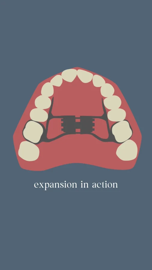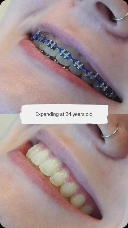

Identify the patients and the measurements
Pirelli and colleagues evaluated 19 children with obstructive sleep apnea and malocclusion before and after expansion. They used CBCT to measure skeletal and airway structures, alongside ENT and sleep-related assessments.
This was a selected clinical group, not a sample of every child who might receive orthodontic care. Before applying the result to another patient, ask whether the starting findings are similar. A study's population helps define the question it can answer and the situations where its conclusions may need more evidence.
Understand what changed on the scans
The researchers reported increased nasal bony width and larger nasal, nasopharyngeal, oropharyngeal, and total upper-airway volumes. These are structural findings beyond the positions of the teeth.
A volume measurement describes the space outlined using the study's imaging method. It does not directly describe every breath a child takes during sleep. For a patient, this distinction matters: “the measured space increased” is a specific finding, while “the child's sleep disorder resolved” requires an assessment of sleep-related outcomes.
Read the design alongside the conclusion
The before-and-after design shows what was observed in the treated group. Without an untreated comparison group, it is harder to separate the intervention's contribution from other changes over time. The small sample and relatively short observation period also limit how broadly the result can be applied.
That does not make the finding unhelpful. It identifies a structural effect worth studying and questions that stronger comparative research can examine. When reading a headline or social caption, look for both the observed result and the limits of the study that produced it. Neither should disappear from the explanation.
Connect the research to an individual decision
Ask the clinician what this paper contributes to your child's assessment. Does the child have a similar orthodontic finding? Which outcome is relevant to the proposed treatment? If a sleep concern is present, how will the team measure it rather than assume an improvement from an image?
Other expansion research has not always demonstrated improved sleep-apnea measurements despite structural change. A balanced plan therefore keeps anatomy and clinical outcomes connected but distinct. The airway evaluation can organize your concerns; the consultation should explain how the child's findings, the evidence, and the follow-up plan fit together.
CBCT describes morphology; the acquisition protocol shapes the measurement
Cone-beam computed tomography provides three-dimensional information about the craniofacial structures included in the scan. For research on airway change, interpretation depends on how the anatomic boundaries are defined and how images before and after treatment are acquired and analyzed. A change in a segmented volume is not necessarily the same as a change in the narrowest clinically relevant passage, and neither directly records airflow during sleep.
The Pirelli study combined anatomical evaluation with other clinical assessments in a selected pediatric sample. Its value is in examining related but distinct outcomes rather than relying on a single photograph. When reading the figures, ask which structures were measured, at which time points, and whether the result concerns skeletal change, nasomaxillary morphology, respiratory testing, or symptoms. These distinctions determine the scope of a defensible conclusion.
Sources: [1]
From a pre–post observation to a causal interpretation
Before-and-after measurements establish that a change was observed during a treatment interval. They do not independently separate the intervention’s effect from growth, measurement variation, or other treatment. Sample size, eligibility criteria, missing follow-up, and the presence or absence of a comparison group therefore matter as much as the appearance of a reconstructed image.
A useful appraisal asks whether all participants were analyzed and whether the investigators prespecified the outcome. It also asks whether the magnitude of change is clinically meaningful, not merely statistically significant. The resulting evidence can support a biologically plausible treatment pathway while leaving uncertainty about who will benefit and by how much. For an individual patient, repeat imaging should answer a clinical question; a research imaging schedule is not an automatic instruction to obtain serial scans solely to document an airway-volume increase.
Bring the conversation to your visit.
- How similar are my child's findings to the children included in this study?
- Which of the reported measurements is relevant to the proposed treatment goal?
- How will any change in sleep or breathing be evaluated separately from imaging?
Supporting studies & resources
These resources add clinical context to the original post. Each publication should be read with its study population, methods, and outcomes in mind.
- Cone beam CT evaluation of skeletal and nasomaxillary complex volume changes after rapid maxillary expansion in OSA children Pirelli et al. · Sleep Medicine · 2021 · Uncontrolled before-and-after study
- Polygraphic evaluation of the effects of different rapid maxillary expansion appliances on sleep quality: A randomized clinical trial Gokce et al. · Sleep and Breathing · 2023 · Randomized trial
- The role of pediatric maxillary expansion on nasal breathing. A systematic review and metanalysis. Calvo-Henriquez et al. · International Journal of Pediatric Otorhinolaryngology · 2020 · Systematic review and meta-analysis