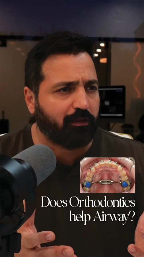
A useful question, with more than one possible answer
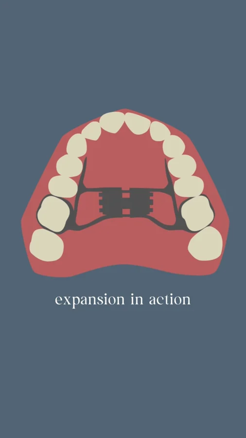
The post asks whether enlarged tonsils and adenoids might sometimes be part of a wider structural picture. That question is worth exploring because an airway assessment should account for the space available and the tissues within it. A narrow palate and enlarged tissues can coexist; finding one does not make the other irrelevant.
The next step is to identify what is actually contributing to symptoms in this patient. A visual impression of the mouth cannot settle what is happening behind the nose or during sleep. Equally, an orthodontic assessment can contribute information that is not captured by looking at tonsil size alone.
What the expansion study contributes
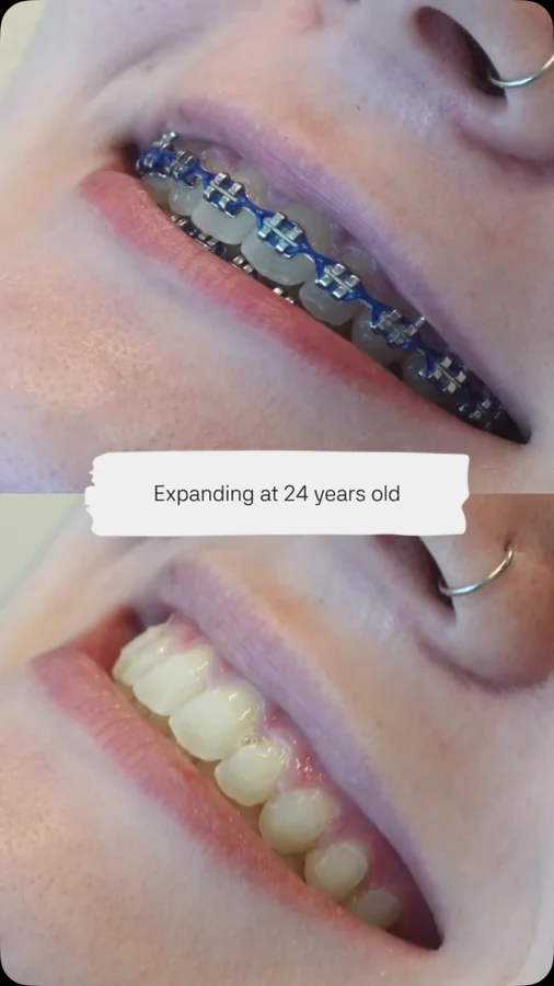
Yoon and colleagues’ 2022 retrospective study compared children with a narrow palate and enlarged tissues who received expansion with a control group. It reported average reductions in adenoid and tonsil volumes in the expansion group. This is a relevant reason to study the relationship between skeletal development and surrounding airway tissues.
The design does not establish that a narrow jaw caused the enlargement, that expansion will shrink every child’s tonsils, or that surgery becomes unnecessary. Those are different questions. The value of the study lies in an observed association in a selected population, rather than a universal treatment rule.
Connect measurements to the reason for treatment
A treatment plan should state its target clearly. Is the aim to correct a transverse jaw deficiency, address nasal obstruction, manage confirmed sleep apnea, or coordinate several of these needs? Each target calls for an appropriate way to assess progress. An imaging measurement and a sleep outcome should not be used interchangeably.
The American Thoracic Society guideline includes orthodontic or dentofacial treatment as a conditional option for selected children with persistent sleep apnea and relevant craniofacial findings. Its cautious evidence rating reinforces the need for selection, collaboration, and follow-up.
Make the consultation a shared conversation
Bring previous ENT evaluations, sleep-study reports, and orthodontic records if you have them. Ask the team to explain how these findings fit together and which question each specialist is answering. This is more useful than asking one treatment to solve every possible source of obstruction.
Families in Northern Virginia, DC, and Maryland can use the airway evaluation to organize their observations before a visit. The goal is an understandable sequence of decisions: what has been found, what still needs investigation, and how the team will know whether the chosen approach has helped.
Lymphoid tissue and transverse skeletal anatomy occupy different diagnostic domains
Adenoids, palatine tonsils, and the maxillary skeleton contribute to different parts of the upper-airway environment. A narrow palate and enlarged lymphoid tissues may coexist, but their coexistence does not establish a single cause or make one treatment a substitute for every other option. Evaluation should describe the structural findings and the respiratory symptoms before assigning a mechanism.
The Yoon cohort provides a specific research observation about tissue-volume changes after expansion in selected children. Its retrospective design does not establish that expansion directly treats every cause of adenotonsillar enlargement or predicts the response of a child outside the studied population. This is a reason to investigate the relationship carefully and coordinate orthodontic and medical assessment, rather than to treat tissue appearance alone as an indication for an expander.
Sources: [1]
Coordinate treatment sequencing and reassessment across specialties
When both a transverse maxillary discrepancy and a medical airway concern are present, the plan should specify the role of each clinician and the sequence of evaluation. A sleep diagnosis, ENT findings, prior treatment, and the orthodontic indication all affect that discussion. The priority is to address the child’s clinically important problem while making the goals of each intervention explicit.
The ATS persistent-OSA guideline is relevant after adenotonsillectomy because it considers remaining contributors, including selected craniofacial findings. It should not be read as a rule that every child needs both procedures or as a reason to delay indicated medical care while awaiting dental change. Follow-up must distinguish tissue dimensions, nasal symptoms, occlusion, and sleep-study outcomes. A reduction in one anatomical measure is informative, but it is not by itself proof that all nocturnal obstruction has been eliminated.
Bring the conversation to your visit.
- Which structures appear to be contributing to these symptoms?
- What outcome would expansion target in this situation?
- How will orthodontic, ENT, and sleep follow-up be coordinated?
Supporting studies & resources
These resources add clinical context to the original post. Each publication should be read with its study population, methods, and outcomes in mind.
- Impact of rapid palatal expansion on the size of adenoids and tonsils in children Yoon et al. · Sleep Medicine · 2022 · Retrospective cohort study
- Management of Persistent, Post-adenotonsillectomy Obstructive Sleep Apnea in Children: An Official American Thoracic Society Clinical Practice Guideline American Thoracic Society · American Journal of Respiratory and Critical Care Medicine · 2024 · Clinical guideline
- Adenotonsillectomy for Snoring and Mild Sleep Apnea in Children: A Randomized Clinical Trial JAMA · 2023 · Randomized trial